II.23 Barley chromosome identification with the C-banding Giemsa stain technique.
K. Noda and K.J. Kasha. Crop Science Dept., University of Guelph, Guelph, Ontario, Canada N1g 2W1.
Chromosome banding provides a new tool for the study of chromosomes by providing a system for their identification. The initial success of such procedures in plants has been limited (Sarma et. al , 1974; Gill and Kimber, 1974) but gradually by modifying procedures they have become available for many species. This report contains a brief outline of procedures we have developed for barley and a description of the banding pattern of the individual chromosomes, as identified by examining trisomic plants.
PROCEDURES
l. Accumulate cells at somatic metaphase by placing root tips (2 to
3 cm in length) in ice-water for 20 hr. in refrigerator.
2. Fix the root-tips with acetic-alcohol (1:3 freshly prepared)
3. Dip the root-tips in tap water.
4. Place the root-tips in 1N HCl at 60°C for 6.5 min. (Exact temperature
is required).
5. Transfer the root-tips to water and prepare slides by squashing
the meristematic tip in a drop of 30% acetic acid under a cover slip.
6. Place the slide against dry ice for freezing and remove the cover
slip.
7. Dry the slides at room temperature, at least for one day.
8. Place the slides in 1N HCl at 60°C for 5 min.
9. Rinse the slides in tap water.
lO. Dry the slides at room temperature for half a day.
11. Immerse the slides in 0.07N NaOH for 35 seconds.
12. Stain in 60X diluted Giemsa solution in Sorensen phosphate buffer
(pH 6.9) for about l hr, monitoring the intensity of stain.
13. Rinse the slides in tap water and dry them at room temperature.
14. Permanently mount the slides in Canada balsam.
Before step 6, it is better to check the cells by a phase contrast microscope to determine if there are well separated chromosomes on the slide. If chromosomes are over-treated and do not show bands, reduce the treatment time of step 8.
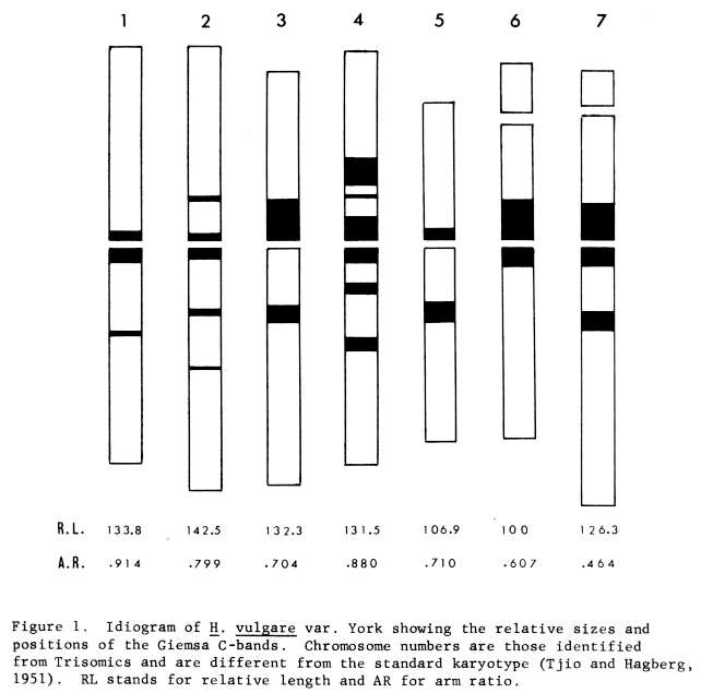
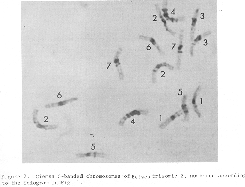
H. vulgare cv. York and the trisomic plants for chromosomes 1, 2, 3 and 4 of cv. Betzes have been examined by the banding technique. Chromosomes 5, 6, and 7 can be readily identified on the basis of chromosome morphology. Almost all bands are located near the centromere regions but the chromosomes can still be identified by their band pattern. In Fig. 1 the pattern of chromosome bands observed in cv. York is illustrated. Fig. 2 shows the banding pattern of Betzes trisomic 2. It is not easy to distinguish chromosome 1 from chromosome 2 since both usually have only small bands and a similar pattern. However, chromosome 1 does not have a clear band in the short arm except the band near the centromere. Chromosome 2 has small bands in both arms in addition to the bands near the centromere region. Chromosome 3 has a large band near the centromere in the short arm but no band near the centromere in the long arm of cv. York (Fig. 1) It does have a band near the middle of the long arm. Chromosome 5, the smallest chromosome has a band pattern similar to 3 except that the centromere band is much smaller. Chromosome 4 is most extensively banded with 2 large bands in the short arm and 3 large bands in the long arm. Chromosomes 6 and 7 can be readily identified by their satellite size and chromosome 7 has a distinct band in the long arm in addition to the centromere banding.
It is important to note that, in contrast to cv. York, chromosome 3 of Betzes (Fig. 2) has a band near the centromere region of chromosome 3. Small bands have also been observed at the secondary constrictions of chromosomes 6 and 7 in some genotypes. Thus, there can be variation in the banding pattern among barley genotypes but the major identifying bands we have described are likely to remain characteristic.
It is also important to note that the chromosomes identified by banding of the trisomics (Fig. 1) appear to be different from the standard idiogram of Tjio and Hagberg (1951). Trisomic 3 appears to be chromosome 1 of Tjio and Hagberg, and Trisomic 1 is their chromosome 3 with a median centromere. Chromosome 2 appears to be the longest among the complement. These results are consistent with the recent data of Tuleen (1973) and Kunzel and Nicoloff (1975), who proposed from studies with chromosomal interchanges that the linkage groups are associated with different chromosomes than the karyotype numbering of Tjio and Hagberg as proposed by Burnham and Hagberg (1956) However, the pretreatments used for root-tips in each study are different and also different from that used by Tjio and Hagberg (1951).
References:
Burnham, C.R, and Hagberg, A. 1956. Cytogenetic notes on chromosomal interchanges in barley. Hereditas 42: 467-482.
Gill, B.S, and Kimber, G, 1974. Giemsa C-banding and the evolution of wheat Proc. Nat. Acad. Sci. USA 71: 4086-4090.
Künzel, G. and Nicoloff, H. 1975. Indications for a necessary reversion of the barley karyogramme. BGN 5: 23-26
Sarma, N.P. and Tanden, S.L. 1974. Banding techniques and plant chromosomes. Current Sci. 43: 635-637.
Tjio, J.H. and Hagberg, A. 1951. Cytological studies on some x-ray mutants of barley. Ann. Aula Dei 2: 149-167.
Tuleen, N.A. 1973. Karyotype analysis of multiple translocation stocks of barley. Can. J. Genet. Cytol. 15: 267-273.