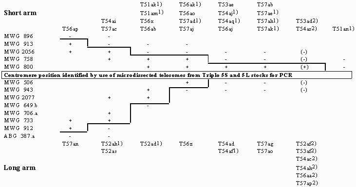
+/-: RFLP probe-specific PCR fragment present/absent. Empty fields: not tested
Using the polymerase chain reaction (PCR) with sequence-specific primers and template DNA from microisolated translocation chromosomes of Vicia faba, Macas et al. (1993) presented a new strategy to physical gene localization in plants. Adaptation of this technique to barley (Sorokin et al. 1994) allowed us to construct for the first time a cytogenetically based physical RFLP map of a barley chromosome. Based on four suitable translocations with breakpoint positions previously localized to relatively short segments of Giemsa-banded somatic metaphase chromosomes (Marthe and Künzel 1994) and an appropriate selection of RFLP probes, the 37 RFLP loci mapped within the Igri/Franka derived linkage group 5 (Graner et al. 1994) could be assigned to five defined subregions of chromosome 5 (Künzel et al. 1995).
Extending the same PCR technique to additional translocation chromosomes, a total of 37 breakpoints have been exactly positioned between the RFLP loci of linkage group 5 (Table 1). With two exceptions, T51an and T57ad, the PCR results fit to the physical data obtained by karyotyping (Table 2). Since the physical position of RFLP loci was confirmed by repeated PCR experiments, the karyotypically determined positions for T1-5am and T5-7ad are erroneous. Based on the PCR results these breakpoint positions were revised.
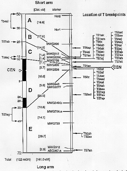
Figure 1 illustrates the location of the 37 breakpoints in relation to the RFLP and the physical map. Most of the breakpoints (31) are clustered within a region of about 15 cM around the centromere. This relatively small part of the genetic map (about 10%) corresponds to about 60% of the total chromosome lenght and represents an area of suppressed meiotic recombination. Exceptionally, one of the few breakpoints located outside this region (T57an) was positioned between the outermost two loci of the long arm.
The localization of a large number of breakpoints exactly between the linear array of genetic markers allowed to refine the breakpoint positions determined previously by karyotype analysis for several translocations. This is especially the case if breakspoints, karyotypically assigned to overlapping segments, occur between the same two flanking markers as specified in Table 2 for 20 of the investigated 37 translocations involving chromosome 5.
Acknowledgement
This research was supported by the Federal Ministry for Research and Technology (Grant No. 0319960B).
Tab. 1. Integration of 37 translocation breakpoints of chromosome 5 into the Igri/Franka derived RFLP linkage map. PCR results with RFLP probe-specific primers and DNA of microisolated translocation chromosomes

+/-: RFLP probe-specific PCR fragment present/absent. Empty fields: not
tested
¹) Only one of the two reciprocally interchanged chromosomes distinct enough for microisolation to be used for PCR
²) Insertion based on breakpoint positions determined by karyotyping (Marthe and Künzel 1994)
Tab. 2. Cytological localization of breakpoints in 20 reciprocal translocations as improved by ordering the breakpoints of chromosome 5 into the linear range of RFLP markers of linkage group 5
                          Breakpoints assigned to segments
                                  defined in mGN¹
                          Determined by         Refined by
T-Line     Chromosome     karyotyping          PCR results
__________________________________________________________
T1-5am       T15am        1S:27 to 36            29 to 36
             T51am        5S:23 to 32            25 to 32
T1-5an*      T15an        1S:(27 to 36)           3 to 14
             T51an        5S:(24 to 33)           0 to 11
T3-5ad       T35ad        3L:20 to 27            21 to 27
             T53ad        5S:10 to 17            11 to 17
T3-5ae       T35ae        3L:55 to 82            56 to 77
             T53ae        5S:10 to 37            11 to 32
T4-5af       T45af        4L:36 to 69            36 to 66
             T54af        5L:29 to 62            29 to 59
T4-5ai       T45ai        4L:28 to 40            28 to 36
             T54ai        5S:38 to 50            38 to 46
T4-5aj       T45aj        4L:28 to 53            28 to 35
             T54aj        5S:25 to 50            25 to 32
T4-5aq       T45aq        4S:13 to 14               14
             T54aq        5S:10 to 11               11
T5-6x        T56x         5S:10 to 32            25 to 32
             T65x         6S:19 to 41            34 to 41
T5-6ab       T56ab        5S:10 to 50            25 to 46
             T65ab        6L:22 to 62            37 to 58
T5-6aj       T56aj        5S:10 to 12            11 to 12
             T65aj        6S: 8 to 10             9 to 10
T5-6ak       T56ak        5S:10 to 50            25 to 46
             T65ak        6L:26 to 66            41 to 62
T5-6ao       T56ao        5S:10 to 48            25 to 46
             T65ao        6L:32 to 70            47 to 68
T5-6ap       T56ap        5S:10 to 46            38 to 46
             T65ap        6L:34 to 70            62 to 70
T5-7ac       T57ac        5S:24 to 50            30 to 46
             T75ac        7L:22 to 48            28 to 44
T5-7ad*      T57ad        5S:(11 to 17)          25 to 46
             T75ad        7L:(10 to 16)          24 to 45
T5-7ah       T57ah        5S:10 to 35            11 to 32
             T75ah        7L:35 to 60            36 to 57
T5-7aj       T57aj        5S:19 to 40            25 to 40
             T75aj        7S:22 to 43            28 to 43
T5-7ak       T57ak        5S:12 to 50            12 to 32
             T75ak        7L:22 to 60            22 to 42
T5-7ao       T57ao        5L:29 to 62            29 to 59
T            T75ao        7L:25 to 58            25 to 55
_________________________________________________________
¹ According to Jensen and Linde-Laursen (1992)

Fig. 1. Location of 37 translocation breakpoints in relation to the
physical and the Igri/Franka derived RFLP map for chromosome 5. Left-hand:
Physical map (idiogram) with measurements in milliGeNomes (mGN) according to
Jensen and Linde-Laursen (1992). Breakpoints used for map construction are
marked by arrows; single arrow for the precisely located breakpoint of T57ab,
cramped arrows indicate the segments within which the breakpoints of T54ai,
T56aj and T57ag are located. Right-hand: Skeletal genetic map indicating the
location of breakpoints within the linear array of RFLP markers. Corresponding
regions of the genetic and physical map are indicated by lines.
References
Graner, A., E. Bauer, A. Kellermann, S. Kirchner, J.K. Muraya, A. Jahoor, and G. Wenzel. 1994. Progress of RFLP-map construction in winter barley. BGN 23: 53-59.
Jensen, J., and I. Linde-Laursen. 1992. Statistical evaluation of length measurements on barley chromosomes with a proposal for a new nomenclature for symbols and positions of cytological markers. Hereditas 117: 51-59.
Künzel, G., A. Sorokin, and F. Marthe. 1995. Progress in relating genetic to physical distances of marker sequences on microisolated barley chromosomes. In: "Kew Chromosome Conference IV", Proc. 4th Kew Chromosome Conference, submitted.
Macas, J., W. Weschke, H. Bäumlein, U. Pich, A. Houben, U. Wobus, and I. Schubert. 1993. Localization of vicilin genes via polymerase chain reaction on microisolated field bean chromosomes. Plant J. 3: 883-886.
Marthe, F., and G. Künzel. 1994. Localization of translocation breakpoints in somatic metaphase chromosomes of barley. Theor. Appl. Genet. 89: 240-248.
Sorokin, A., F. Marthe, A. Houben, U. Pich, A. Graner, and G. Künzel. 1994. Polymerase chain reaction mediated localization of RFLP clones to microisolated translocation chromosomes of barley. Genome 37: 500-555.